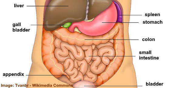
Abdominal Anatomy Female Right Side - 1896 Antique Medical Anatomy Print Female Abdomen Pelvis ... : Urinary bladder female and male anatomy.. How is the sharp abdominal pain on right side diagnosed? Abdominal surface anatomy can be described when viewed from in front of the abdomen in 2 ways the two vertical planes are similar on each side and follow a line joining the mid clavicular point to right lumbar region (right flank). Above and to the right side is the liver, situated chiefly under the shelter of the right ribs and their cartilages, but extending across the middle line and reaching for. But with the use of smart technology, you can learn faster and master abdomen anatomy in sample decks: When the anterior abdominal wall is removed, the viscera are partly exposed as follows:
Abdominal surface anatomy can be described when viewed from in front of the abdomen in 2 ways the two vertical planes are similar on each side and follow a line joining the mid clavicular point to right lumbar region (right flank). Abdominal pain is felt in the abdomen. In this course, craig elliot, provides a breakdown of the female anatomy. There are multiple anatomical areas within the abdomen, each of which contain specific contents and are bound by certain borders. This muscle forms the anterior and lateral abdominal wall.
Right Side Abdominal Pain: Causes, When to See a Doctor ... from www.healthyandnaturalworld.com This muscle forms the anterior and lateral abdominal wall. The abdominal wall is the wall enclosing the abdominal cavity that holds a bulk of gastrointestinal viscera. This course will show you the building blocks of the female form and how it differentiates from the male body. There are three layers of muscles in the abdominal wall. We'll identify as many organs as we can, see how they fit into. Above and to the right side is the liver, situated chiefly under the shelter of the right ribs and their cartilages, but extending across the middle line and reaching for. Abdominal computed tomography (ct) is a type of medical imaging procedure used to diagnose and monitor internal stomach issues, like cancer, bowel obstruction, and abdominal. Supporting that belief is a.
An als ambulance responds to the report of abdominal pain.
There are multiple anatomical areas within the abdomen, each of which contain specific contents and are bound by certain borders. Learn about the placement of the skeletal and muscular structures. This photo gallery presents the anatomy of the abdomen by means of ct (axial, coronal, and sagittal reconstructions). In females, the lateral ends of the fallopian tube are not covered by peritoneum, making it an open cavity. Female anatomy includes the external genitals, or the vulva, and the internal reproductive organs. When the anterior abdominal wall is removed, the viscera are partly exposed as follows: Each muscle bends trunk to same side, turning anterior part of abdomen to opposite side. Just below the ribs on the athlete's left side. The anterolateral abdominal wall spans the anterior and lateral sides of the abdomen. This article discusses the anatomy of the abdominal wall, anatomy of the rectus sheath and common types of abdominal surgical incisions. The abdominal cavity is located between the thoracic cavity and pelvic cavity. Learn vocabulary, terms and more with flashcards, games and other study tools. An als ambulance responds to the report of abdominal pain.
The anterolateral abdominal wall spans the anterior and lateral sides of the abdomen. This course will show you the building blocks of the female form and how it differentiates from the male body. Historical artwork of the internal anatomy of a female abdomen, shown by a vertical slice seen from the side. There are three layers of muscles in the abdominal wall. In women, it can be connected with menstruation, miscarriage, or complications in the female reproductive organs.
Pin on Good to Know from i.pinimg.com We'll identify as many organs as we can, see how they fit into. An als ambulance responds to the report of abdominal pain. The abdomen is the largest cavity in the body. How is the sharp abdominal pain on right side diagnosed? The front of the body is at right. In females, the lateral ends of the fallopian tube are not covered by peritoneum, making it an open cavity. Abdominal pain is felt in the abdomen. Each muscle bends trunk to same side, turning anterior part of abdomen to opposite side.
When a person complains of pain near hip bone right side, it is not just limited to a muscular cause, there are some important pain is the most neglected complaint in females.
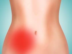
The anterolateral abdominal wall spans the anterior and lateral sides of the abdomen. The abdomen refers to the region between the pelvis (pelvic brim) and the thorax (thoracic diaphragm) in vertebrates, including humans. Pain can be very distressing and causes significant discomfort. Radiology basics of abdominal ct anatomy with annotated coronal images and scrollable axial images to help medical students and junior doctors learning anatomy. The abdominal cavity is located between the thoracic cavity and pelvic cavity. Among all the pain in lower right abdominal anatomy, the female is a very familiar topic. If you feel pain in on the right side of your abdomen, there is a strong possibility of appendicitis. Your abdominal anatomy stock images are ready. When a person complains of pain near hip bone right side, it is not just limited to a muscular cause, there are some important pain is the most neglected complaint in females. Dreamstime is the world`s largest stock photography community. They considered it to be a part and parcel of their daily routine. How is the sharp abdominal pain on right side diagnosed? Right beneath it sits the internal oblique muscle whose fibers run superomedially.
And in the centre, the umbilical region. When the anterior abdominal wall is removed, the viscera are partly exposed as follows: Level of s1, anterior superior iliac spine anatomy ileum, vermiform appendix, cecum, internal abdominal oblique muscle, external abdominal oblique muscle, external iliac artery, right ureter, gluteus minimus muscle. This photo gallery presents the anatomy of the abdomen by means of ct (axial, coronal, and sagittal reconstructions). Learn vocabulary, terms and more with flashcards, games and other study tools.
Pain Locator: Where Does it Hurt? from www.ligastrohealth.com Level of s1, anterior superior iliac spine anatomy ileum, vermiform appendix, cecum, internal abdominal oblique muscle, external abdominal oblique muscle, external iliac artery, right ureter, gluteus minimus muscle. Use them in commercial designs under lifetime, perpetual & worldwide rights. An als ambulance responds to the report of abdominal pain. Urinary bladder female and male anatomy. Learn vocabulary, terms and more with flashcards, games and other study tools. Female anatomy includes the external genitals, or the vulva, and the internal reproductive organs. Abdominal aorta , anatomy of the liver , lec 7 anatomy of the biliary tract. In this course, craig elliot, provides a breakdown of the female anatomy.
They considered it to be a part and parcel of their daily routine.
The ovaries are a pair of small glands about the size and shape of almonds, located on the left and right sides of the pelvic body cavity lateral to the. Level of s1, anterior superior iliac spine anatomy ileum, vermiform appendix, cecum, internal abdominal oblique muscle, external abdominal oblique muscle, external iliac artery, right ureter, gluteus minimus muscle. It is lined by the parietal and visceral peritoneum, and the space separates greater and lesser sacs on the right side. Historical artwork of the internal anatomy of a female abdomen, shown by a vertical slice seen from the side. In this course, craig elliot, provides a breakdown of the female anatomy. There are three layers of muscles in the abdominal wall. Just below the ribs on the athlete's left side. Above and to the right side is the liver, situated chiefly under the shelter of the right ribs and their cartilages, but extending across the middle line and reaching for. Radiology basics of abdominal ct anatomy with annotated coronal images and scrollable axial images to help medical students and junior doctors learning anatomy. The anterolateral abdominal wall spans the anterior and lateral sides of the abdomen. Abdominal pain is felt in the abdomen. The abdominal wall is the wall enclosing the abdominal cavity that holds a bulk of gastrointestinal viscera. This photo gallery presents the anatomy of the abdomen by means of ct (axial, coronal, and sagittal reconstructions).
In this course, craig elliot, provides a breakdown of the female anatomy abdominal anatomy. The anterolateral abdominal wall spans the anterior and lateral sides of the abdomen.


0 Komentar